

Imaging changes in children and adolescents diagnosed with autism spectrum disorder: A literature review

Imaging changes in children and adolescents diagnosed with autism spectrum disorder: A literature review

Imaging changes in children and adolescents diagnosed with autism spectrum disorder: A literature review

Citation: Vargas-Díaz M, Martínez-Núñez KM, Amador-Rodero EM, Rebolledo-Cobos RC. Cambios imagenológicos en niños y adolescentes diagnosticados con trastorno del espectro autista: Revisión de la literatura. Univ Salud [Internet]. 2026; 28(3):e10172. DOI: 10.22267/rus.262803.10172.


# ABSTRACT

Introduction: Autism spectrum disorder (ASD) is a neurodevelopmental disorder characterized by difficulties in social interaction and communication, as well as repetitive behaviors. Despite its high prevalence, the pathophysiological basis of ASD is not yet fully understood. Objective: To analyze findings from neuroimaging studies that provide evidence of pathophysiological changes in the brains of individuals with ASD. Materials and methods: A literature review was conducted in PubMed, UpToDate, ClinicalKey, Google Scholar, and ScienceDirect between February and August 2024. Selected elements of the PRISMA statement were considered to organize the search and study-selection process. Results: The reviewed studies identified structural differences in the amygdala, cerebellum, and other cortical regions, together with alterations in neural connectivity and atypical patterns of brain activation. These dysfunctions are associated with difficulties in social interaction and sensory integration. Conclusion: Neuroimaging techniques provide valuable information on brain alterations associated with ASD and may contribute to earlier diagnosis and the development of more effective therapeutic interventions.

Keywords: Autism spectrum disorder; Children; Neuroimaging. (Source: DeCS, Bireme).

Objetivo de desenvolvimento sustentável: Health and well-being. (Source: SDG, WHO).


# RESUMEN

Introducción: El trastorno del espectro autista (TEA) es un trastorno del neurodesarrollo caracterizado por dificultades en la interacción social, comunicación y comportamientos repetitivos. A pesar de su alta prevalencia, las bases fisiopatológicas del TEA no se comprenden completamente. Objetivo: Analizar los hallazgos de estudios de neuroimagen que evidencian cambios fisiopatológicos en el cerebro de personas con TEA. Materiales y métodos: Se realizó una revisión de la literatura en las bases de datos PubMed, UpToDate, ClinicalKey, Google Académico y ScienceDirect entre febrero y agosto de 2024. Para la organización del proceso de búsqueda y selección de los artículos se tuvieron en cuenta algunos lineamientos de la guía PRISMA. Resultados: Los estudios revisados evidenciaron diferencias estructurales en la amígdala, el cerebelo y otras áreas corticales, así como alteraciones en la conectividad neuronal y patrones atípicos de activación cerebral. Estas disfunciones se asocian con dificultades en la interacción social y la integración sensorial. Conclusión: Las técnicas de neuroimagen proporcionan información valiosa sobre las alteraciones cerebrales asociadas con el TEA y pueden contribuir al diagnóstico temprano y al desarrollo de intervenciones terapéuticas más efectivas.

Palabras clave: Trastorno del espectro autista; Niños; Neuroimagen. (Fuente: DeCS, Bireme).

Objetivo de desarrollo sostenible: Salud y bienestar. (Fuente: ODS, OMS).


# RESUMO

Introdução: O transtorno do espectro autista (TEA) é um transtorno do neurodesenvolvimento caracterizado por dificuldades na interação social, comunicação e comportamentos repetitivos. Apesar de sua alta prevalência, a base fisiopatológica do TEA não é totalmente compreendida. Objetivo: Analisar os achados de estudos de neuroimagem que demonstram alterações fisiopatológicas no cérebro de indivíduos com TEA. Materiais e métodos: Foi realizada uma revisão da literatura nas bases de dados PubMed, UpToDate, ClinicalKey, Google Scholar e ScienceDirect entre fevereiro e agosto de 2024. As diretrizes PRISMA foram utilizadas para organizar a busca e o processo de seleção dos artigos. Resultados: Os estudos revisados ​​revelaram diferenças estruturais na amígdala, no cerebelo e em outras áreas corticais, bem como alterações na conectividade neuronal e padrões atípicos de ativação cerebral. Essas disfunções estão associadas a dificuldades na interação social e na integração sensorial. Conclusão: As técnicas de neuroimagem fornecem informações valiosas sobre as alterações cerebrais associadas ao TEA e podem contribuir para o diagnóstico precoce e o desenvolvimento de intervenções terapêuticas mais eficazes.

Palavras-chave: Transtorno do espectro autista; Crianças; Neuroimagem. (Source: DeCS, Bireme).

Objetivo de desenvolvimento sustentável: Saúde e bem-estar. (Source: SDG, WHO).

# INTRODUCTION

Autism spectrum disorder (ASD) is a neurodevelopmental disorder characterized primarily by difficulties in social interaction, impairments in verbal and nonverbal communication, and the presence of repetitive patterns of behavior or restricted interests. These manifestations typically emerge in early childhood and may persist throughout life, producing varying degrees of impairment in social, cognitive, and behavioral functioning1,2.

The Diagnostic and Statistical Manual of Mental Disorders, Fifth Edition (DSM-5), supports the preceding definition and further establishes that these manifestations must be present from the early developmental period and cause clinically significant impairment in daily functioning3.

Symptoms generally emerge in early childhood and persist throughout life. Diagnosis is confirmed using standardized diagnostic protocols and structured clinical instruments, with neuroimaging techniques such as magnetic resonance imaging supporting the understanding of their neurobiological basis4. These techniques have broadened knowledge of the neurobiological mechanisms associated with ASD. Magnetic resonance imaging (MRI), positron emission tomography (PET), single-photon emission computed tomography (SPECT), and magnetoencephalography (MEG) have provided relevant evidence regarding structural and functional alterations in the brains of individuals with this disorder5.

Magnetic resonance imaging studies have identified variations in the size and function of several brain structures, including the cerebellum and amygdala, which in some cases exhibit volumetric differences in individuals with ASD6. Likewise, functional magnetic resonance imaging has demonstrated distinct patterns of brain activation during tasks involving language, decision-making, and social interaction7. Diffusion magnetic resonance imaging has also enabled examination of connectivity among different brain regions, revealing potential alterations in neural networks involved in communication and sensory integration processes8.

Positron emission tomography studies have enabled assessment of cerebral metabolic activity and have shown differences in glucose utilization in specific brain regions in individuals with ASD. In addition, alterations have been identified in neurotransmitters such as serotonin and gamma-aminobutyric acid (GABA), both of which play fundamental roles in neuronal communication and the regulation of multiple cognitive and social functions9.

Other approaches, including single-photon emission computed tomography and magnetoencephalography, have also contributed relevant evidence to the study of ASD. SPECT has identified atypical cerebral blood-flow patterns in regions involved in emotional regulation and social interaction, whereas MEG has demonstrated potential alterations in the synchronization of brain activity during tasks involving sensory processing and social perception10.

Despite the growing body of research in this field, findings concerning pathophysiological alterations at the neuroanatomical and neurophysiological levels in ASD remain dispersed across multiple studies. Accordingly, a literature review integrating and critically analyzing the principal available scientific findings on these alterations is warranted. Therefore, the present study aimed to analyze the existing scientific evidence on the main neuroanatomical and neurophysiological pathophysiological abnormalities associated with ASD, thereby contributing to a more comprehensive understanding of the neurobiological mechanisms involved in this disorder.


# MATERIALS AND METHODS

# Study design

Literature review with a descriptive approach.

A review of the scientific literature was conducted to address the PICO question (Population: children with autism spectrum disorder; Intervention/Exposure: neuroimaging studies or brain imaging assessment; Comparison: neurotypical children without ASD; Outcome: neuroimaging changes): What imaging changes are observed in children and adolescents diagnosed with autism spectrum disorder?

# Selection criteria and search strategy

The literature review was conducted between February and April 2024. The following databases were used: PubMed, UpToDate, ClinicalKey, Google Scholar, and ScienceDirect.

The search strategy used the Boolean operators AND and OR and combined the terms Autism, Pathophysiology, Children, and Neuroimaging. The search equation was: ("Autism Spectrum Disorder" OR "Autism") AND ("Neuroanatomy" OR "Neurophysiology" OR "Brain alterations"). For PubMed, filters were applied for study type, language, publication date, and document type in order to select relevant, high-quality studies. In Google Scholar, the following DeCS terms were entered into the search engine: Trastorno del Espectro Autisa or Autismo; Trastorno del espectro autista y neuroanatomía; Trastorno del espectro autista y neurofisiología or neuroanatomía. In ClinicalKey, the following equation was entered: ("autism spectrum disorder" OR autism OR "autistic disorder" AND child* OR pediatric* AND neuroimaging OR "brain imaging"). In UpToDate, a topic search was conducted using "Autism spectrum disorder children neuroimaging".

# Selection criteria

Inclusion criteria: Scientific articles focused on children and adolescents with a primary diagnosis of autism spectrum disorder (ASD); systematic reviews with or without meta-analysis addressing neuroanatomical or neurophysiological alterations associated with the disorder and describing neuroimaging techniques used to identify structural or functional brain changes. Articles published in English or Spanish and studies with clearly described methodologies were also considered.

Exclusion criteria: Articles addressing ASD secondary to underlying pathological conditions were excluded.

# Data extraction

The preselected articles underwent critical appraisal to verify their relevance, methodological quality, and alignment with the study objective. Inclusion and exclusion criteria were established a priori to minimize potential bias during evidence selection.

The information sources comprised academic databases and indexed scientific literature, thereby supporting greater validity and methodological rigor among the included studies. Subsequently, data were systematically extracted from the selected articles for analysis.

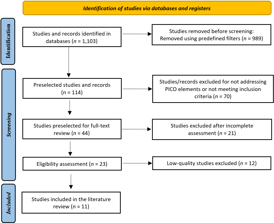
The literature identification, selection, and analysis process was organized with reference to the phases proposed in the PRISMA (Preferred Reporting Items for Systematic Reviews and Meta-Analyses) statement, including identification, screening, eligibility, and inclusion. This approach enhanced transparency in evidence selection and reduced potential bias during the review process (Figure 1).

# Figure 1. Flow diagram of the search, classification, and selection of articles
<b>Figure 1.</b>.  Flow diagram of the search, classification, and selection of articles

    # RESULTS

    The initial search identified a total of 1,103 articles (PubMed, 642; UpToDate, 102; ClinicalKey, 115; Google Scholar, 244). After application of the predefined filters, 114 articles remained for analysis (PubMed, 37; UpToDate, 28; ClinicalKey, 16; Google Scholar, 33). Of these, 70 were excluded for failing to meet the inclusion criteria, leaving 44 articles for methodological appraisal. Systematic reviews and meta-analyses were evaluated using PRISMA guidance, whereas observational studies were assessed using the STROBE statement. Following this appraisal, 23 articles were preselected, and 11 studies were ultimately included in the present review after consideration of their level of evidence and grade of recommendation (Table 1).

    # Synthesis of results

    This review identified multiple neurobiological alterations and specific clinical phenotypes associated with autism spectrum disorder and other neurological conditions that affect cognitive functioning and brain connectivity. The included studies provide a broad perspective on variations in brain structure and connectivity-related function in autism spectrum disorder and other related neurological conditions.

    # Functional alterations

    One of the included studies applied neuroimaging techniques (EEG, fMRI, fNIRS, MRI) in 551 high-risk (HR) and low-risk (LR) infants to investigate structural and functional brain differences, with particular emphasis on responses to auditory, visual, and social stimuli11. Among HR infants who were subsequently diagnosed with ASD (HR+), reduced gray matter volume was observed, a feature associated with cognitive function and information processing.

    # Brain connectivity

    Functional connectivity was investigated using EEG and MEG in a sample of 52 individuals12. Long-range underconnectivity was identified in interhemispheric and anteroposterior connections between the frontal and occipital cortices, suggesting impaired information integration in individuals with ASD13.

    Similarly, another study examined brain activity during memory tasks in individuals with autism spectrum disorder and neurotypical participants. Participants with ASD exhibited reduced prefrontal activation accompanied by greater activation in parietal and temporal regions, suggesting a possible cerebral reorganization to accommodate the cognitive demands of memory processing14.

    # Structural alterations

    Neuroanatomical characteristics, including cortical thickness, surface area, and cortical volume, were also examined using magnetic resonance imaging to compare autistic and neurotypical individuals15.

    The findings showed increased cortical thickness in frontal, temporal, parietal, and occipital regions during childhood in autistic individuals, followed by accelerated cortical thinning during adolescence, highlighting differences in neuroanatomical development.

    Positron emission tomography (PET) was used to examine the availability of mitochondrial electron transport chain complex I in 48 patients with ASD. The findings demonstrated reduced availability of this complex in several brain regions, including the anterior cingulate cortex, superior temporal gyrus, and dorsolateral prefrontal cortex, which may indicate altered cerebral energy metabolism16.

    Another study investigated cortical responses to vibrotactile stimuli in 24 individuals with autism spectrum disorder and neurotypical participants using EEG and MEG17. The findings suggest that EEG-derived inter-trial coherence (ITC) may represent a useful biomarker for detecting differences in sensory processing in individuals with autism spectrum disorder18.

    A systematic review and meta-analysis involving 3,514 individuals examined anatomical and functional alterations in ASD. Reduced functional activity was identified in key brain regions, including the insula and anterior cingulate/medial frontal cortex, together with alterations in the default mode network and motor and sensory regions, indicating substantial changes in brain function and structure19.

    An analysis of 4,831 individuals identified differences in structural and functional abnormalities between attention-deficit/hyperactivity disorder (ADHD) and ASD20. ADHD was characterized by reduced gray matter volume in the ventromedial orbitofrontal region, whereas ASD showed increases in frontotemporal regions. Functionally, ASD was associated with hypoactivation of the medial prefrontal cortex and hyperactivation patterns in the bilateral ventrolateral prefrontal cortices, indicating functional alterations distinct from those observed in ADHD. Together with evidence of atypical trajectories of brain maturation, these findings suggest that the structural and functional alterations associated with ASD may vary according to developmental stage21,22.

    Several studies have suggested that sex-related brain differences in ASD may be associated with neurobiological processes that interact with developmental trajectories. Multimodal neuroimaging research integrated with neurogenetic evidence has identified potential sex-related differences in the relationship between neuroanatomy and ASD, whereas subsequent systematic reviews have indicated that these differences may vary according to age and developmental stage, with possible involvement of genetic and endocrine mechanisms17,23. However, the available evidence regarding sex-related differences in ASD brain circuits and networks remains limited and has not identified consistent female-specific patterns.

    # Metabolic findings

    Another analysis used magnetic resonance spectroscopy (MRS) to quantify brain metabolites and found lower concentrations of GABA and NAA in individuals with ASD, suggesting an excitation/inhibition imbalance and alterations in neuronal integrity, particularly in limbic regions and in children24.

    Finally, gray matter differences were compared between individuals with anorexia nervosa (AN) and ASD. Although the two disorders share certain behavioral similarities, no consistent neuroanatomical correlates were identified between AN and ASD, suggesting that behavioral similarities do not necessarily reflect shared brain structures25.

    The reviewed studies highlight both unique and shared patterns of neurological alteration in ASD, ranging from differences in gray matter volume and functional connectivity to variations in mitochondrial complex availability and cerebral metabolites. These findings underscore the neurobiological complexity of autism spectrum disorder and the importance of continued research into specific and differential biomarkers that may improve early diagnosis and individualized treatment in this population.

    # Table 1. Characteristics of the studies included in the literature review
    Author Design Population Intervention Comparison Outcome
    (P) (I) (C) (O)
    Ayoub MJ, Keegan L, Tager-Flusberg H, Gill SV5 Systematic review N = 551 Neuroimaging studies (EEG, fMRI, fNIRS, MRI) used to identify structural and functional differences between HR and LR infants, focusing on brain activation in response to auditory, visual, and social stimuli HR infants who subsequently received an ASD diagnosis (HR+), HR infants who were not diagnosed with ASD (HR-), and LR infants Reduced gray matter volume, associated with cognitive function and information processing
    O'Reilly C, Lewis JD, Elsabbagh M12 Systematic review N = 52 Electroencephalography (EEG) and magnetoencephalography (MEG) recordings used to assess functional connectivity Not applicable
    Desaunay P, Guillery B, Moussaoui E, Eustache F, Bowler DM, Guénolé F7 Systematic review N = 33 Functional neuroimaging studies assessing memory function in individuals with autism spectrum disorder and typically developing individuals Comparisons of brain activity and performance on memory tasks between groups of individuals with autism spectrum disorder and typically developing individuals Reduced prefrontal activation, compensated by greater parietal or temporal activation
    Pretzsch CM, Ecker C6 Literature review N = 35 Analysis of neuroanatomical characteristics, including cortical thickness, surface area, cortical volume, local gyrification index, and gray-white matter tissue contrast using magnetic resonance imaging Comparison of autistic and neurotypical individuals, considering developmental trajectories of neuroanatomical characteristics across the lifespan Cortical thickness increases in frontal, temporal, parietal, and occipital regions during childhood, followed by accelerated thinning during adolescence
    Kato Y, Yokokura M, Iwabuchi T, Murayama C, Harada T, Goto T, et al15 Observational study N = 48 Assessment of mitochondrial electron transport chain complex I (MC-I) availability using positron emission tomography (PET) Mitochondrial electron availability in the brains of individuals with ASD versus typically developing individuals Reduced availability of mitochondrial complex I in frontal, temporal, occipital, thalamic, and motor brain regions
    Ahlfors SP, Graham S, Alho J, et al10 Observational study N = 24 Assessment of differences in sensory processing between individuals with autism spectrum disorder and neurotypical individuals using electroencephalography (EEG) and a vibrotactile stimulation paradigm Comparison between individuals with autism spectrum disorder and neurotypical individuals EEG inter-trial coherence may serve as a biomarker of altered sensory processing in individuals with autism spectrum disorder
    Guo, Z., Tang, X., Xiao, S. et al11 Systematic review with meta-analysis N = 3,514 To explore the most consistent findings on functional and structural brain alterations in individuals with autism spectrum disorder (ASD) from resting-state functional imaging and voxel-based morphometry (VBM) studies Individuals with autism spectrum disorder (ASD) and typically developing (TD) individuals Structural and functional brain alterations, with reduced activity in the insula, anterior cingulate/medial frontal cortex, and motor, sensory, and default mode networks
    Lukito S, Norman L, Carlisi C, Radua J, Hart H, Simonoff E, Rubia K16 N = 4,831 To identify structural and functional abnormalities that distinguish and overlap between individuals with attention-deficit/hyperactivity disorder (ADHD) and autism spectrum disorder (ASD) Individuals with attention-deficit/hyperactivity disorder (ADHD) and those with autism spectrum disorder (ASD) ASD is characterized by increases in frontotemporal regions and medial prefrontal hypoactivation, whereas ADHD shows bilateral ventrolateral prefrontal hyperactivation
    Walsh MJM, Wallace GL, Gallegos SM, Braden BB17 Systematic review N = 50 To understand how sex-related biological factors, such as hormones and genes, influence neurodevelopmental trajectories in females with ASD Females with autism spectrum disorder (ASD) and neurotypical cohorts, as well as different age groups within the ASD population Females with ASD exhibit distinctive neurodevelopmental trajectories influenced by sex-related biological factors, although the brain circuits involved remain understudied
    Thomson AR, Pasanta D, Arichi T, Puts NA9 Systematic review with meta-analysis N = 35 Application of proton magnetic resonance spectroscopy (1H-MRS) to quantify brain metabolite concentrations in individuals with autism spectrum disorder Brain metabolite concentrations in individuals with autism spectrum disorder and individuals without the disorder Reduced GABA and NAA, associated with excitation/inhibition imbalance and altered neuronal integrity, particularly in children and limbic regions
    Sader M, Williams JHG, Waiter GD18 Meta-analysis N = 69 Analysis of structural magnetic resonance imaging literature to identify shared structural neural correlates in anorexia nervosa (AN) and autism spectrum disorder (ASD) Structural brain alterations in individuals with anorexia nervosa and autism spectrum disorder No consistent neuroanatomical correlates were identified between anorexia nervosa and autism spectrum disorder despite behavioral similarities in food selectivity

      # DISCUSSION

      The review of neuroimaging studies in autism spectrum disorder (ASD) highlights substantial progress in identifying structural and functional biomarkers, thereby enabling a more comprehensive understanding of the underlying neurological alterations11. Nevertheless, comparison of these findings with the broader scientific literature reveals important discrepancies that reflect the complexity and heterogeneity of the disorder. Whereas some studies report reductions in gray matter volume, others describe opposing patterns depending on age and the characteristics of the population studied.

      In this regard, previous studies have reported alterations in long-range brain connectivity associated with difficulties in information integration15. However, research by Eric Courchesne and colleagues has demonstrated early increases in brain volume in children with ASD26. This discrepancy suggests that structural changes may follow a nonlinear trajectory, characterized initially by accelerated growth that may subsequently stabilize or even decline.

      With respect to specific brain structures, magnetic resonance imaging studies have demonstrated variations in cerebellar and amygdala size27. Although the present review reports enlargement of these structures, other studies indicate that such changes are not constant across development. For example, Catherine Lord has proposed that these regions may be enlarged during early developmental stages and subsequently show patterns of normalization or reduction28. This observation underscores the importance of considering developmental stage when interpreting neuroimaging findings.

      From a physiological perspective, functional magnetic resonance imaging has identified differences in brain activation during tasks involving language, decision-making, and social interaction13. These alterations suggest a distinct functional organization in individuals with ASD, with direct implications for cognitive and social performance. However, variability across findings indicates that brain regions are not affected uniformly in all individuals.

      Regarding brain connectivity, some studies support a disconnection hypothesis involving distant brain regions, which may impair the integration of complex information13. Nevertheless, the model proposed by Nancy Minshew suggests that, in addition to long-range hypoconnectivity, local hyperconnectivity may also be present29. This dual pattern may help explain why some individuals with ASD exhibit highly developed specific abilities alongside difficulties in more global functions.

      Metabolic studies using positron emission tomography have also demonstrated alterations in cerebral glucose utilization30. These differences in metabolic activity may be associated with impairments in the regulation of cognitive and social functions. However, there is no consensus as to whether these alterations represent a direct cause of the disorder or a consequence of adaptive brain processes.

      Within this framework, Geraldine Dawson has proposed that some neurobiological changes may be interpreted as compensatory mechanisms developed by the brain in response to ASD-related challenges31. This perspective introduces a more dynamic view of the disorder in which the brain exhibits not only deficits but also adaptive strategies.

      Another relevant consideration is the role of neurotransmitters in ASD. Imbalances have been identified in systems involving serotonin and GABA, both of which participate in the regulation of mood, behavior, and sensory processing18. These alterations may contribute to characteristic manifestations of the disorder, including behavioral rigidity and difficulties in social interaction.

      Sex-related differences have also been reported in the manifestation of ASD in males and females17. The "extreme male brain" theory proposed by Simon Baron-Cohen suggests that the disorder is associated with a more systemizing cognitive profile32. However, this theory has been challenged by more recent research emphasizing social camouflaging in females, which may contribute to underdiagnosis in this population.

      Likewise, comparisons between ASD and other disorders such as ADHD allow both similarities and differences in neurobiological patterns to be identified16. Although shared features are present, including impairments in executive functions, important differences in brain organization have also been observed. In this context, Russell Barkley has noted that the two disorders may overlap in attentional and inhibitory-control processes, which can complicate clinical differentiation33.

      Despite these advances, variability in the findings remains a major limitation of neuroimaging research in ASD23. Differences in methodological designs, sample sizes, and the techniques employed hinder comparisons across studies and limit the generalizability of the findings.

      # Limitations

      The present review has several limitations. First, heterogeneity in methodological designs, neuroimaging techniques, and characteristics of the studied populations limits direct comparison of the findings. Second, the inclusion of studies with different levels of evidence may introduce bias into the interpretation of the results.


      # CONCLUSIONS

      The reviewed evidence demonstrates that ASD is associated with multiple neuroanatomical and functional alterations, including structural changes in brain regions such as the amygdala, cerebellum, and cortical areas; alterations in connectivity and brain activation; and changes in metabolism and sensory processing.

      These findings underscore the complexity and heterogeneity of the neurobiological basis of ASD and its potential relationship with difficulties in social interaction, communication, and sensory integration. Neuroimaging techniques are valuable tools for understanding these changes and advancing the identification of potential biomarkers; however, the available evidence remains heterogeneous across studies and does not support a single neuroimaging pattern characteristic of ASD.

      Longitudinal studies with large samples and homogeneous methodological criteria are needed to determine the trajectory of these alterations and their potential utility in supporting early diagnosis and guiding individualized interventions.

      Within this context, physiotherapy has an important role in integrating neurobiological evidence with clinical reasoning and functional intervention. Understanding neuroimaging changes can support physiotherapists in designing more specific and individualized therapeutic strategies aimed at promoting neuroplasticity, sensorimotor self-regulation, and functional coordination.

      In summary, neuroimaging research contributes to a broader understanding of the neurobiological basis of ASD and provides relevant evidence to strengthen physiotherapy practice grounded in knowledge of neurological, sensory, and functional characteristics. Integration between neuroscience and physiotherapy offers an opportunity to consolidate interdisciplinary models of care that connect an understanding of brain mechanisms with the functional needs of individuals with ASD, with the potential to enhance participation, well-being, and quality of life while supporting their families.


      # Acknowledgments

      The authors thank Universidad Libre de Colombia for providing research time and bibliographic resources. Artificial intelligence tools were used to support the development of this work, particularly the organization, structuring, and refinement of the written content; their use helped improve the clarity and coherence of the manuscript and facilitated a more organized and comprehensible presentation of the information. Nevertheless, the analysis, selection of information, and conclusions presented herein remain the responsibility of the authors.


      # REFERENCES

      1. 1. Lord C, Brugha TS, Charman T, Cusack J, Dumas G, Frazier T, et al. Autism spectrum disorder. Nat Rev Dis Primers [Internet]. 2020; 6(1):5. DOI: https://doi.org/10.1038/s41572-019-0138-4
      2. 2. Arberas C, Ruggieri V. Autismo: Aspectos genéticos y biológicos. Medicina [Internet]. 2019 [citado 2026 Abr 12]; 79(Suppl 1):16-21. Disponible en: https://www.scielo.org.ar/scielo.php?script=sci_arttext&pid=S0025-76802019000200005&lng=es
      3. 3. American Psychiatric Association. Diagnostic and statistical manual of mental disorders: DSM-5. 5th ed. Washington (USA): American Psychiatric Association; 2013. Disponible en: https://www.appi.org/dsm
      4. 4. Schielen SJC, Pilmeyer J, Aldenkamp AP, Zinger S. The diagnosis of ASD with MRI: a systematic review and meta-analysis. Transl Psychiatry [Internet]. 2024; 14(1):318. DOI: https://doi.org/10.1038/s41398-024-03024-5
      5. 5. Ayoub MJ, Keegan L, Tager-Flusberg H, Gill SV. Neuroimaging techniques as descriptive and diagnostic tools for infants at risk for autism spectrum disorder: a systematic review. Brain Sci [Interent]. 2022; 12(5):602. DOI: https://doi.org/10.3390/brainsci12050602
      6. 6. Pretzsch CM, Ecker C. Structural neuroimaging phenotypes and associated molecular and genomic underpinnings in autism: a review. Front Neurosci [Internet]. 2023; 17:1172779. DOI: https://doi.org/10.3389/fnins.2023.1172779
      7. 7. Desaunay P, Guillery B, Moussaoui E, Eustache F, Bowler DM, Guénolé F. Brain correlates of declarative memory atypicalities in autism: a systematic review of functional neuroimaging findings. Mol Autism [Internet]. 2023; 14(1):2. Disponible en: https://link.springer.com/article/10.1186/s13229-022-00525-2
      8. 8. Vidal Barraza NV. Análisis de imágenes de resonancia magnética por difusión en adolescentes con autismo [Tesis]. Concepción (CHL): Universidad de Concepción; 2023. Disponible en: https://repositorio.udec.cl/items/faec1bdd-4cf4-4e26-a5e7-497f1731f32f
      9. 9. Thomson AR, Pasanta D, Arichi T, Puts NAP. Neurometabolite differences in autism as assessed with magnetic resonance spectroscopy: A systematic review and meta-analysis. Neurosci Biobehav Rev [Internet]. 2024; 162:105728. DOI: https://doi.org/10.1016/j.neubiorev.2024.105728
      10. 10. Ahlfors SP, Graham S, Alho J, Joseph RM, McGuiggan NM, Nayal Z, et al. Magnetoencephalography and electroencephalography can both detect differences in cortical responses to vibrotactile stimuli in individuals on the autism spectrum. Front Psychiatry [Internet]. 2022; 13:902332. DOI: https://doi.org/10.3389/fpsyt.2022.902332
      11. 11. Guo Z, Tang X, Xiao S, Yan H, Sun S, Yan Z, et al. Systematic review and meta-analysis: multimodal functional and anatomical neural alterations in autism spectrum disorder. Molecular Autism [Internet]. 2024; 15(1):16. DOI: https://doi.org/10.1186/s13229-024-00593-6
      12. 12. O'Reilly C, Lewis JD, Elsabbagh M. Is functional brain connectivity atypical in autism? A systematic review of EEG and MEG studies. PLoS One [Internet]. 2017; 12(5):e0175870. DOI: https://doi.org/10.1371/journal.pone.0175870
      13. 13. Echeverry CD, Moreno GLJ. Bases del conocimiento molecular del autismo con variantes VARS y NRXN1: a propósito de un caso. IJEPH [Internet]. 2023; 6(1):e8637. DOI: https://doi.org/10.18041/2665-427X/ijeph.1.8637
      14. 14. Martinez-Morga M, Paz Quesada M, Bueno C, Martinez Salvador S. Bases neurobiológicas del autismo y modelos celulares para su estudio experimental. Medicina [Internet]. 2019; 79(Suppl 1):27-32. Disponible en: https://www.scielo.org.ar/scielo.php?script=sci_arttext&pid=S0025-76802019000200007
      15. 15. Kato Y, Yokokura M, Iwabuchi T, Murayama C, Harada T, Goto T, et al. Lower availability of mitochondrial complex I in anterior cingulate cortex in autism: a positron emission tomography study. Am J Psychiatry [Interent]. 2023; 180(4):277-284. DOI: https://doi.org/10.1176/appi.ajp.22010014
      16. 16. Lukito S, Norman L, Carlisi C, Radua J, Hart H, Simonoff E, et al. Comparative meta-analyses of brain structural and functional abnormalities during cognitive control in attention-deficit/hyperactivity disorder and autism spectrum disorder. Psychol Med [Internet]. 2020; 50(6):894-919. DOI: https://doi.org/10.1017/S0033291720000574
      17. 17. Walsh MJM, Wallace GL, Gallegos SM, Braden BB. Brain-based sex differences in autism spectrum disorder across the lifespan: A systematic review of structural MRI, fMRI, and DTI findings. Neuroimage Clin [Internet]. 2021; 31:102719. DOI: https://doi.org/10.1016/j.nicl.2021.102719
      18. 18. Sader M, Williams JHG, Waiter GD. A meta-analytic investigation of grey matter differences in anorexia nervosa and autism spectrum disorder. Eur Eat Disord Rev [Internet]. 2022; 30(5):560-579. DOI: https://doi.org/10.1002/erv.2915
      19. 19. Hazlett HC, Gu H, Munsell BC, Kim SH, Styner M, Wolff JJ, et al. Early brain development in infants at high risk for autism spectrum disorder. Nature [Internet]. 2017; 542(7641):348-351. DOI: https://doi.org/10.1038/nature21369
      20. 20. Di Martino A, O'Connor D, Chen B, Alaerts K, Anderson JS, Assaf M, et al. Enhancing studies of the connectome in autism using the autism brain imaging data exchange II. Sci Data [Internet]. 2017; 4(1):170010. DOI: https://doi.org/10.1038/sdata.2017.10
      21. 21. Wang Z, Zheng L, Yang L, Yin S, Yu S, Smith R, et al. Structural and functional whole brain changes in autism spectrum disorder at different age stages. Eur Child Adolesc Psychiatry [Internet]. 2024; 34(5):1589-1602. DOI: https://doi.org/10.1007/s00787-024-02585-6
      22. 22. Ecker C, Bookheimer SY, Murphy DGM. Neuroimaging in autism spectrum disorder: brain structure and function across the lifespan. Lancet Neurol [Internet]. 2015; 14(11):1121-1134. DOI: https://doi.org/10.1016/S1474-4422(15)00050-2
      23. 23. Floris DL, Filho JOA, Lai MC, Giavasis S, Oldehinkel M, Mennes M, et al. Towards robust and replicable sex differences in the intrinsic brain function of autism. Mol Autism [Internet]. 2021; 12(1):19. DOI: https://doi.org/10.1016/S1474-4422(15)00050-2
      24. 24. Chen C, Van Horn JD, GENDAAR Research Consortium. Developmental neurogenetics and multimodal neuroimaging of sex differences in autism. Brain Imaging Behav [Internet]. 2017; 11(1):38-61. DOI: https://doi.org/10.1007/s11682-015-9504-3
      25. 25. Hazlett HC, Poe MD, Gerig G, Styner M, Chappell C, Smith R, et al. Early brain overgrowth in autism associated with an increase in cortical surface area before age 2 years. Arch Gen Psychiatry [Internet]. 2011; 68(5):467-476. DOI: https://doi.org/10.1001/archgenpsychiatry.2011.39
      26. 26. Courchesne E, Campbell K, Solso S. Brain growth across the life span in autism: age-specific changes in anatomical pathology. Brain Res [Internet]. 2011; 1380:138-145. DOI: https://doi.org/10.1016/j.brainres.2010.09.101
      27. 27. Rogel-Ortiz FJ. Autismo. Gac Med Mex [Internet]. 2005 Abr [citado 2026 Ago 10]; 141(2):143-147. Disponible en: https://www.scielo.org.mx/scielo.php?script=sci_arttext&pid=S0016-38132005000200009&lng=es
      28. 28. Lord C, Elsabbagh M, Baird G, Veenstra-VanderWeele J. Autism spectrum disorder. Lancet [Internet]. 2018; 392(10146):508-520. DOI: https://doi.org/10.1016/S0140-6736(18)31129-2
      29. 29. Minshew NJ, Williams DL. The new neurobiology of autism: cortex, connectivity, and neuronal organization. Arch Neurol [Internet]. 2007; 64(7):945-950. DOI: https://doi.org/10.1001/archneur.64.7.945
      30. 30. Palau Baduell M. Análisis de las alteraciones magnetoencefalográficas en pacientes con trastornos del espectro autista [Tesis]. Barcelona (ESP): Universitat Autònoma de Barcelona; 2017. Disponible en: https://dialnet.unirioja.es/servlet/tesis?codigo=139238
      31. 31. Dawson G, Rogers S, Munson J, Smith M, Winter J, Greenson J, et al. Randomized, controlled trial of an intervention for toddlers with autism: the Early Start Denver Model. Pediatrics [Internet]. 2010; 125(1):e17-e23. DOI: https://doi.org/10.1542/peds.2009-0958
      32. 32. Baron-Cohen S. The extreme male brain theory of autism. Trends Cogn Sci [Internet]. 2002; 6(6):248-254. DOI: https://doi.org/10.1016/S1364-6613(02)01904-6
      33. 33. Barkley RA. Attention-deficit hyperactivity disorder: A handbook for diagnosis and treatment. 4th ed. New York (USA): Guilford Press; 2014. Disponible en: https://www.guilford.com/books/Attention-Deficit-Hyperactivity-Disorder/Russell-Barkley/9781462538874